The global landscape of zoonotic pathogens requires continuous surveillance to mitigate biological threats to human health. Rabies, a lethal viral encephalomyelitis affecting all mammalian species, remains a primary focus of international biosecurity frameworks. In the United Kingdom, terrestrial mammals have maintained freedom from classical rabies virus since 1922. However, the presence of specific lyssaviruses within the domestic bat population ensures that biological risk persists across both rural counties and the Greater London metropolitan area.
- What is the Clinical Definition of Bat Rabies?
- Which Bat Species Carry Lyssaviruses in the United Kingdom?
- How Does the Lyssavirus Transmit from Bats to Humans?
- What Happens Inside the Human Body Post-Exposure?
- What Clinical Symptoms Indicate Active Infection?
- What Immediate Medical Protocol Follows a Bat Exposure Event?
- How Does the United Kingdom Monitor and Regulate Bat Rabies?
- What Historical Milestones Define Our Understanding of This Disease?
- What are the Ecological and Future Health Implications of Bat Rabies?
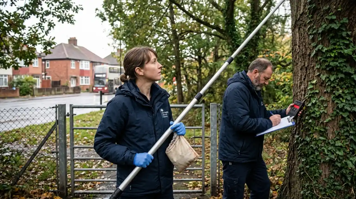
Understanding the interface between human populations and chiropteran vectors is critical for preventing exposure events in densely populated urban centers. The Animal and Plant Health Agency tracks viral prevalence within wild populations to inform national health directives, monitoring habitats extending from remote woodlands into London parklands. Through systematic monitoring and targeted vaccination strategies, the UK Health Security Agency maintains robust defenses against localized outbreaks. This analysis establishes the operational boundaries, virological attributes, and emergency response protocols concerning the bat rabies entity.
What is the Clinical Definition of Bat Rabies?
Bat rabies refers to an acute central nervous system infection caused by RNA viruses belonging to the genus Lyssavirus, carried by chiropteran hosts, which causes fatal encephalomyelitis in humans and other mammals once clinical symptoms manifest.
The causative agents of bat rabies belong to the family Rhabdoviridae. While terrestrial animals typically transmit classical rabies virus, insectivorous bats in Europe carry distinct species known as European Bat Lyssavirus Type 1 and European Bat Lyssavirus Type 2. These pathogens are negative-stranded, single-stranded RNA viruses enveloped in a lipid bilayer containing surface glycoproteins. The virus targets the peripheral nervous system before utilizing retrograde axonal transport to invade the brain and spinal cord.
The World Health Organization classifies all lyssaviruses as agents capable of inducing clinical rabies. Consequently, public health definitions—enforced strictly across London hospitals and regional clinics—treat exposures to bat lyssaviruses with the same clinical urgency as terrestrial rabies. The infection exhibits a variable incubation period, which typically spans three to twelve weeks in humans but can extend for years depending on the inoculation site and viral load. Once the virus crosses the blood-brain barrier, it induces profound inflammation of the gray matter, leading to uniform mortality without rapid medical intervention.
Which Bat Species Carry Lyssaviruses in the United Kingdom?
The Daubenton’s bat acts as the primary reservoir for European Bat Lyssavirus Type 2, whereas the serotine bat serves as the primary host for European Bat Lyssavirus Type 1 within Great Britain.
The ecology of European bat lyssaviruses relies on specific host selection across distinct geographic ranges, including urban ecosystems like the London green belt. Passive surveillance data compiled by the Animal and Plant Health Agency indicates that more than 21,000 bats have undergone diagnostic testing since 1986. The overwhelming majority of confirmed positive cases in the United Kingdom involve two specific insectivorous species:
- Daubenton’s Bat (Myotis daubentoni): This species prefers riparian habitats, utilizing rivers, canals, and lakes—such as the River Thames and urban London waterways—for foraging. It serves as the primary reservoir for European Bat Lyssavirus Type 2, with cases identified across England, Scotland, and Wales.
- Serotine Bat (Eptesicus serotinus): This species is larger, typically roosting in older buildings and foraging over pastures, urban parklands, and woodland edges. It serves as the exclusive host for European Bat Lyssavirus Type 1 within the UK, with detections concentrated primarily in southern regions, including counties bordering Greater London.
While the UK boasts 18 resident bat species, classical pipistrelles, brown long-eared bats, and noctules do not historically harbor these specific viral strains. Isolated detections in other species represent anomalous spillover events rather than sustained reservoir establishment. The prevalence rate remains exceedingly low, affecting less than one percent of the wild population tested via the national passive screening program.
How Does the Lyssavirus Transmit from Bats to Humans?
Transmission occurs when infectious saliva containing the virus enters the human body via a bite or scratch from a bat, or through direct contact between the animal’s salivary secretions and open wounds or mucous membranes.
The mechanical process of viral transmission depends entirely on the introduction of viable viral particles into deep tissue or vascular pathways. The lyssavirus replication cycle concentrates heavy viral loads inside the salivary glands of the chiropteran host during the infectious phase. When an infected animal inflicts a transcutaneous injury, the virus bypasses the protective epidermal layer of the human host. Because British insectivorous bats possess microscopic, needle-like teeth, a transmission-grade bite can occur without causing significant pain or visible hemorrhaging, a risk factor that London public health campaigns emphasize for people clearing out household lofts.
Alternative transmission pathways involve the exposure of broken skin or mucous membranes to fresh saliva. If an individual handles an injured bat without dermal protection, salivary transfer into existing lacerations can initiate infection. Aerogenic transmission within roosting caves represents a theoretical risk due to highly concentrated viral aerosols, but this pathway remains undocumented outside of dense colonial settings in the Americas. The virus cannot penetrate intact, healthy skin, nor does it survive long-term exposure to desiccation, ultraviolet radiation, or chemical detergents outside the host organism.
What Happens Inside the Human Body Post-Exposure?
The virus replicates locally within muscle tissue before binding to nicotinic acetylcholine receptors at neuromuscular junctions, initiating an ascending journey through the peripheral nerves to the central nervous system.
The pathophysiology of lyssavirus infection follows a strict neurotropic trajectory designed to evade host immune defenses. Following the initial inoculation event, the virus undergoes a localized eclipse phase within the muscle cells adjacent to the wound site. During this early period, the pathogen remains largely undetectable by circulating lymphocytes, preventing the activation of adaptive immune responses. The virus utilizes its surface glycoproteins to lock onto specific cellular receptors located at the neuromuscular junction:
- Nicotinic Acetylcholine Receptor (nAChR): The primary site of cellular entry for the virus at the motor endplate.
- Neural Cell Adhesion Molecule (NCAM): A secondary receptor facilitating cellular attachment within the nervous tissue.
- P75 Neurotrophin Receptor (p75NTR): A structural protein involved in guiding internal viral transport dynamics.
Once inside the peripheral axon, the virus moves toward the central nervous system via retrograde axoplasmic transport. This movement occurs at an estimated velocity of 50 to 100 millimeters per day. The speed of progression dictates the length of the incubation period; bites on the face or neck lead to accelerated central nervous system invasion compared to injuries sustained on the lower extremities. Upon reaching the spinal cord dorsal root ganglia, the virus multiplies rapidly and ascends into the brainstem, cerebellum, and cerebral cortex, causing irreversible neuronal dysfunction.
What Clinical Symptoms Indicate Active Infection?
Initial symptoms manifest as localized paresthesia, fever, and headache, which rapidly advance into severe neurological signs including hydrophobia, aerophobia, generalized muscle spasms, acute agitation, and terminal flaccid paralysis.
The clinical manifestation of encephalomyelitic lyssavirus disease follows a predictable progression divided into prodromal, acute neurological, and comatose phases. The prodromal stage lasts between two and ten days, presenting with non-specific constitutional signs. The most reliable early diagnostic sign is localized paresthesia—characterized by tingling, burning, or numbness—at the exact site of the original, healed bat bite. This sensory disturbance stems from viral replication within the local sensory ganglion cells.
As the virus spreads throughout the brain matter, the patient enters the acute neurological phase, which displays two distinct clinical typologies:
Encephalitic Rabies
This form occurs in approximately eighty percent of human cases. The primary clinical sign is hydrophobia, where attempts to swallow liquids trigger violent, involuntary spasms of the diaphragm, pharyngeal, and laryngeal muscles. This reaction is caused by viral destruction of the brainstem nuclei that regulate swallowing, coupled with an exaggerated reflex response to sensory stimuli. Aerophobia, where a gentle breeze on the skin induces identical spasms, occurs concurrently. Patients experience hyper-reactivity, confusion, hallucinations, and fluctuating consciousness.
Paralytic Rabies
This alternative presentation accounts for twenty percent of cases. It avoids the dramatic spastic episodes of the encephalitic form, presenting instead with progressive muscle weakness that begins at the bitten limb and spreads symmetrically. The clinical presentation mimics Guillain-Barré syndrome, which historically complicates differential diagnoses in major London neurological centers. Both clinical forms ultimately converge on widespread central nervous system destruction, culminating in respiratory failure, vascular collapse, coma, and death within days of neurological onset.
What Immediate Medical Protocol Follows a Bat Exposure Event?
The mandatory medical protocol requires immediate mechanical washing of the wound with soap and water for fifteen minutes, followed by the urgent administration of post-exposure prophylaxis via rabies vaccines and human rabies immunoglobulin.
Managing a potential lyssavirus exposure requires immediate action to neutralize the virus before it gains access to the peripheral nervous system. The physical removal of viral particles from the wound site represents the most critical step in minimizing viral load. Rinsing the site under running water with a standard surfactant breaks down the delicate lipid envelope of the virus, rendering it non-infectious. Health guidelines emphasize that individuals must not close the wound with sutures unless absolutely necessary, as mechanical closure can force viral particles deeper into the tissue beds.
Following localized decontamination, patients must immediately contact emergency medical services via NHS 111 or an urgent general practitioner appointment. The UK Health Security Agency manages a dedicated Rabies and Immunoglobulin Service based in London to guide clinical risk assessments nationwide. If the service determines that an exposure occurred, it authorizes the immediate distribution of post-exposure prophylaxis to local emergency rooms:
- Naïve Individuals (Unvaccinated): A primary course consists of four separate doses of an inactivated rabies vaccine administered intramuscularly into the deltoid muscle on days 0, 3, 7, and 21.
- Human Rabies Immunoglobulin (HRIG): Infiltrated directly into and around the margins of the wound on day 0, providing immediate passive antibodies to neutralize the virus before the vaccine stimulates active immunity.
- Previously Vaccinated Individuals: A simplified course consisting of two vaccine booster doses administered on days 0 and 3, omitting the requirement for human rabies immunoglobulin.
How Does the United Kingdom Monitor and Regulate Bat Rabies?
The state manages risk via a national passive surveillance program led by the Animal and Plant Health Agency, statutory notifiable disease reporting laws, and strict biosecurity regulations governing international animal transit.
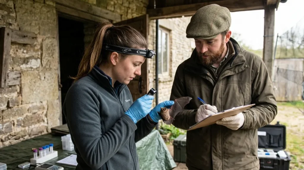
The long-term mitigation of lyssavirus threats relies on a comprehensive legal and scientific infrastructure. Because the United Kingdom maintains a statutory designation of freedom from classical terrestrial rabies, any detection of lyssavirus within domestic borders triggers immediate multi-agency response protocols. Under the Rabies (Control) Order 1974, rabies is classified as a notifiable animal disease. Veterinarians, wildlife rehabilitators, and members of the public are legally required to report any suspected cases or unexplained bat mortalities to the Department for Environment, Food and Rural Affairs.
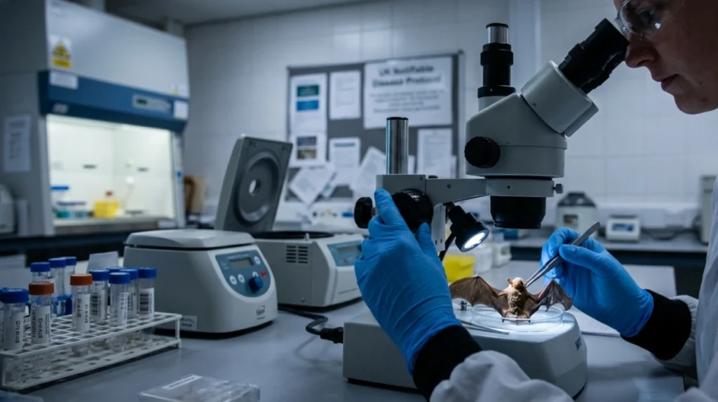
The foundational monitoring tool is the passive surveillance framework managed by the Animal and Plant Health Agency at its Weybridge reference laboratory, located just outside Greater London. The laboratory collects dead bats submitted by the public and wildlife organizations to perform fluorescent antibody testing on brain tissue smears. This diagnostic test allows pathologists to visualize lyssavirus antigens under ultraviolet microscopy.
To prevent the introduction of classical rabies virus into the domestic ecosystem via major transit hubs like London Heathrow Airport, the country enforces the Non-Commercial Movement of Pet Animals Order, alongside the long-standing Pet Travel Scheme. These regulatory frameworks require valid microchipping, mandatory rabies vaccinations, and specific waiting periods for all domestic companion animals entering the British Isles from non-exempt international locations.
What Historical Milestones Define Our Understanding of This Disease?
Key historical milestones include the 1954 isolation of the first bat lyssavirus in Germany, the 1985 death of a Finnish researcher that initiated European surveillance, and the 2002 Scottish fatality that confirmed domestic endemicity.
The scientific characterization of bat-borne lyssaviruses developed independently from the classical understanding of canine rabies. For centuries, public health strategies focused exclusively on domestic dogs and red foxes as the sole vectors of the disease. This paradigm shifted in 1954 when researchers in Hamburg, Germany, successfully isolated a non-classical lyssavirus from a wild bat. This discovery proved that chiropterans could host distinct lineages of the virus without displaying the obvious furious symptoms seen in terrestrial mammals.
The contemporary framework for European bat conservation and public health interaction was shaped by two critical human fatalities:
- The 1985 Finland Case: A veteran bat researcher contracted and succumbed to a European Bat Lyssavirus Type 2 infection. This event forced European nations, including public health authorities in London, to establish formal active and passive surveillance programs to track the virus across the continent.
- The 2002 United Kingdom Case: A bat conservationist in Scotland died after contracting European Bat Lyssavirus Type 2 from a Daubenton’s bat bite. This represented the first indigenous human rabies fatality in Great Britain since 1902, demonstrating conclusively that the pathogen was endemic within the local bat population.
These milestones shifted public policy away from general eradication efforts toward targeted education, pre-exposure vaccination protocols for licensed bat handlers, and strict handling guidelines enforced by conservation bodies like the London Bat Group and the Bat Conservation Trust.
What are the Ecological and Future Health Implications of Bat Rabies?
Climate shifts and expanding urban footprints alter bat roosting distributions, creating new interfaces for public interaction that require long-term environmental protection laws and advanced pan-lyssavirus vaccine research.
The interaction between human populations and chiropteran habitats is changing due to climate patterns and expanding urban developments. As temperatures rise across Northern Europe, the geographic range of specific host species shifts, altering the distribution of European Bat Lyssavirus Type 1 and Type 2. Urbanization forces bats to seek roosting alternatives in residential roofs, soffits, and commercial structures, increasing the likelihood of accidental human contact within the dense, built environment of London.
Future public health strategies must balance disease management with environmental conservation. Bats provide vital ecosystem services, including insect population control and urban agricultural pest reduction. Consequently, eradication programs are ecologically counterproductive and legally prohibited under the Wildlife and Countryside Act 1981, which grants full protection to all British bat species and their roosts, regardless of whether they are located in rural woodlands or Central London developments.
To address the evolving risk, medical research centers focus on developing next-generation, pan-lyssavirus vaccines. Current human vaccines rely on inactivated classical rabies virus strains, which offer robust cross-protection against European Bat Lyssavirus Type 1 and Type 2 but show reduced efficacy against divergent global lineages, such as the West Caucasian bat lyssavirus. Advancements in viral vector and mRNA vaccine technologies aim to produce universal immunotherapies. These developments ensure that The Londoner News readers and the global population remain fully protected against all present and emerging lyssavirus variants.
Does the UK have rabies?
The UK is free of classical rabies virus in terrestrial mammals. However, rare bat-associated lyssaviruses (European Bat Lyssavirus Type 1 and Type 2) are present in a very small number of wild bats.